Diagnostic imaging of canine abdomen using radiography, ultrasonography and endoscopy
|

 |
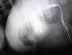
Laparoscopic-guided and laparotomy assisted intraoperative mesenteric portography and ultrasound-guided percutaneous splenoportography were found to be easily executable procedures to evaluate the hepatic blood flow in clinical settings and to bring the future clinical cases of portosystemic shunts in diagnostic ambit.
Capnoperitoneography using CO2 at8mmof Hg intrabdominal pressurethrough automatic insufflator was an easy procedure while exercising accurate control and monitoring over the intrabdominal pressure. It enhanced the visceral visualization of abdominal organs in general and was particularly useful in the evaluation of liver lobes and its borders.
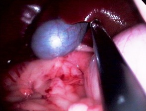
Laparoscopy at 11 mm of Hg provided adequate 3D-visualization of the abdominal organs. It is a better executable procedure in comparison to exploratory laparotomy. Visualization of shape, size, colour, peripheral blood circulation and presence of minute lesions on visceral surface of liver, spleen, stomach, kidneys, intestine, uterus, ovary, urinary bladder was possible with laparoscopy.
The combine usage of all the three diagnostic imaging modalities helped in converging at definitive diagnosis, thus, enabling appropriate and precise intervention at the earliest.
Ultrasonography of bovine abdominal cavity
Scanning area for ultrasonographic examinations of bovine abdominal organs
Organs |
Scanning area |
Reticulum |
Abdominal/thoracic region at the level of elbow in left 7th to 5th ICSs and right 7th to 6th
ICS as well as in median, right paramedian and left paramedian regions as per Braun and Gotz (1994) |
| |
Omasum |
11th to 6th ICSs dorsoventrally as per Braun and Blessing (2006) |
| |
Abomasum |
Ventral abdominal region caudal to xiphoid process as per Braun et al. (1997) |
| |
Rumen |
In left flank and 12th ICS as per Tschour et al. (2008) |
| |
Spleen |
12th to 6th ICSs as per Braun et al. (2008) |
| |
Liver |
12th to 6th ICSs as per Braun (1990)
|
| |
Gall bladder |
12th to 9th ICSs and paracostal area as per Braun (1990) |
| |
Pancreas |
Behind last rib and 12th ICS as per Pusterla et al. (1997) |
| |
Small intestine |
12th to 8th ICSs and right ventral flank as described by Braun et al. (1995) |
| |
Large intestine |
12th ICS and right dorsal flank as per Braun et al. (2001) |
| |
Kidneys |
Just behind the last rib, dorsal aspect of last ICS and 1st to 3rd lumbar spaces on right side |
| |


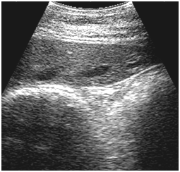
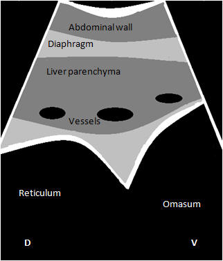
Ultrasonogram of cow showing reticulum, omasum, and liver from the right 6th ICS
Bovine Ultrasonography
Ultrasonography of bovine reticulum can be done from 7th to 5th ICS in left side, and 6th to 5th ICS in right side.
Omasum can be seen as an amotile echogenic semicircular arc from 11th to 6th ICS.
Small intestines can be scanned in ventral half of right abdomen and it is difficult to locate them cranial to 11th ICS.
Bull’s eye or sandwich like appearances of intestinal loops are characteristic of intestinal intussusceptions and can be observed transabdominally.
Cecum and SLAC are observed in dorsal to mid-flank region and differentiation between cecum and PLAC, and SLAC and descending colon is not possible.
Liver can be scanned from 12th to 6th ICS as an isoechogenic structure and the gall bladder as anechoic pear shaped structure from 12th ICS to 9th ICS and ventral to costal arch.
There are no specific diagnostic ultrasonographic features of omasal impaction.
Both right and left kidneys can be viewed from right paralumbar fossa transabdominally.
Dilation of CVC was a characteristic feature in cases of right sided heart failure.


