Anesthesia in yak
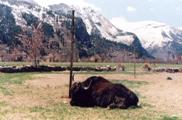
| The Department of Surgery and Radiology, Palampur has pioneered the research in anesthesia in yaks. The Yak belongs to the order Artiodactyla, the sub family bovine of family bovidae. Yak keeping has been an important survival strategy for nomads and mountain agriculturists. The chemical restraint, general and regional anesthetic techniques has been extensively studied for the first time in the Department of Surgery and Radiology, College of Veterinary and Animal Sciences, Palampur. |
| Recommendations for chemical restraint and general anesthesia in yaks |
| Recommendations for regional anesthesia in yak |
1. Cornual nerve block:
The horn of yak was chiefly innervated by the corneal nerve (lacrimal-ophthalmic-trigeminal)which had two distinct branches in temporal fossa. A caudal branch of auriculopalpebral nerve also reached to the base of horn. Eight ml of analgesic is to beinjected mid way between base of horn and lateral canthus of the eye blocks the dorsal and ventral branches of the corneal nerve. The onset of analgesia and return of sension occurrs 7.89 ± 0.54 and 74.55 ± 4.09 minutes respectively after the injection of the analgesic. The analgesia remained for 66.67 ± 3.91 minutes.
2. Auriculopalpebral nerve block:
The auriculopalpebral nerve across from the dorsal buccal nerve of facial nerve. As it reaches in the temporal fossa it forms a superficial temporal plexus. The nerve then sent rami to the upper eye lid at the lateral canthus and to the lower eye lid. Five ml of analgesic injected, blocks the palpebral supply after 7.89 ± 0.54 minutes of the injection resulting into akinesia of the lower eye lid and hypokinesia of the upper eye lid towards lateral canthus. the motor reflex remains blocked for 125.55 ± 4.12 minutes. The blinking of the eye lids resums after 132.33 ± 4.84 minutes.
3. Petersons nevre block:
The motor supply to the muscles of eye ball came through the oculomotor, trochlear and abducent nerves. Ophthalmic and maxillary divisions of the trigeminal nerve which enters through the foramen orbitorotundum furnishes sensory innervations to them. Fifteen ml of the analgesic deposited close to the foramen orbitorotundum by penetrating the needle between the vertical ramus of the mandible (caudally) and supraorbital process of the zygomatic arch (rostrally) fixes the eye ball (movement) and produces complete desensitization after 11.71 ± 0.75 minutes of injection. The analgesia remaines for 85 to 110 minutes.
4. Paravertebral nerve block:
The flank area in yak was innervated by the last two thoracic (t-13, t-14) and first two lumber (L-1, L-2) spinal nerves. four ml of the analgesic infiltrated at the origin of nerve superficially and 5 ml deeply blocks the respective branches of flank area. The onset of analgesia occurred at 2.55 ± 0.24 minutes in upper flank and 4.00 ± 0.33 minutes in the lower flank. The duration of analgesia was 112.22 ± 4.50 and 56.67 ± 3.12 minutes respectively in these areas. Rumenotomy can successfully be performed through upper flank incision.
5. Lumbar epidural block:
The yak has 14 thoracic and 5 lumber vertebrae. The lumber spines are straight. the interaricual space is significant and approachable from dorsal side. The first interlumber space was chosen for the lumber epidural analgesia. Seven ml of analgesic isintroduced. the analgesia was induced within 13.28 ± 0.92 minutes in upper flank and 16.43 ± 0.84 minutes in lower flank bilaterally with return of sensation in 99.71 ± 4.15 minutes and 91,43 ± 3.48 minutes respectively post injection. The duration of analgesia remaines for 75.00 ± 3.27 minutes and 86.43 ± 3.73 minutes in the lower flank and para lumber fossa respectively. Laparotomy can be performed through left flank successfully.
6. Caudal epidural nerve block:
The spinal cord terminats at 2nd sacral vertebra and a large interarcual gap occurrs between the saccrococcygeal and first inter-coccygeal articulations. It provides the ideal site for caudal epidural analgesia. Four ml of the analgesic is deposited in the first intercoccygeal space for obtaining the epidural analgesia. The analgesic effect appeares in tail and perineum within 1 to 3 minutes of the injection, which persists for 110 ± 3.02, 52 ± 4.08 and 77.2 ± 2.78 minutes at the base of the tail, tip of the tail and perineum respectively.
7. Brachial plexus block:
The brachial plexus of yak is formed by union of the ventral branches of C-6, C-7, C-8, T-1 and T-2 spinal nerves. The nerve roots exit through the scalene and forming the plexus in synsarcosis. eleven nerves arise from the plexus which innervates the thoracic limb and ventral and lateral parts of the thoracic wall. The roots of the brachial plexus were approached from the point of the scapulo-humeral joint. The needle is inserted 7 to 8 cm straight between the shoulder and the chest wall and 40 ml of the analgesic was deposited to block the middle, dorsal and ventral roots of the plexus. The onset of the analgesia was observed 12.57 ± 0.92, 16.00 ± 1.00 and 16.86 ± 0.80 minutes in the antebrachium, metacarpal and digital regions. the analgesia remains for 39 ± 2.54, 35.00 ± 2.44 and 32.14 ± 1.84 minutes respectively in these regions.
8. Ring block of metacarpus and metatarsus:
The manus region in yak is innervated by the branches of ulnar, median and radial nerves and the pes region was innervated by the tibial, fibular, saphenoius and posterior cutaneous nerves. the ring block is made at the proximal one third of metacarpus/metatarsus by infiltrating 20ml of analgesic under the skin as far as bone forming a ring. The onset of analgesia was observed 4.33 ± 0.41 and 6.00 ± 0.41 minutes, duration of analgesia 78.89 ± 3.61 and 71.11 ± 3.09 minutes and return of sensation 83.22 ± 3.68 and 77.00 ± 3.14 minutes after the injection of the analgesic in metacarpal and digital regions respectively. In the hind limb, the analgesia was induced in 4.22 ± 0.36 and 5.67 ± 0.44 minutes, and remained for 80.00 ± 4.33 and 68 ± 3.89 minutes in the metatarsal and digital regions respectively.
Anesthesia in Neonatal Calves
Anesthesia in Neonatal Calves |
| S.No. | Name of the preanaesthetics/sedative/tranquillizer/ Surgical anaesthetic combination | Dose rate | Route of administration | Type of Effect |
1 | Atropine sulfate | 0.04 mg/kg | Subcutaneously | Anticholinergic |
2 | Xylazine hydrochloride | 0.22 mg/kg | Intramuscularly | Sedation |
3 | Diazepam | 0.3 mg/kg | Intravenously | Tranquillization |
4 | Triflupromazine hydrochloride | 0.5 mg/kg | Intravenously | Tranquillization |
5 | Acepromazine maleate | 0.75 mg/kg | Intravenously | Tranquillization |
6 | Detomidine hydrochloride | 0.02 mg/kg | Intramuscularly | Sedation |
7 | Chloral hydrate, 4% solution | 7.5gm/100kg | Intravenously | Narcosis |
8 | Chloral hydrate +magnesium sulfate (Chloral-mag), 1:1, 6% solution | 10gm/100kg | Intravenously | Narcosis |
| 9 | Chloral hydrate +magnesium sulfate (Chloral-mag), 1:1, 6% solution 10 min later, Thiopentone sodium, 5% | 10gm/100kg 15mg/kg'to effect' | Intravenously Intravenously | General anaesthesia |
| 10 | Xylazine hydrochloride plus Ketamine hydrochloride | 0.22mg/kg single 5mg/kg syringe | Intramuscularly | Balanced surgical anaesthesia |
| 11 | Detomidine hydrochloride plus Ketamine hydrochloride | 0.02 mg/kg single 7.5 mg/kg syringe | Intramuscularly | Balanced surgical anaesthesia |
| 12 | Medetomidine hydrochloride | 0.01 mg/kg | Intramuscularly | Sedation |
| 13 | Atropine sulfate 10 min later, Medetomidine hydrochloride plus Ketamine hydrochloride | 0.04 mg/kg 0.015mg/kg single syringe 10mg/kg | Subcutaneously Intramuscularly | Balanced surgical anaesthesia |
| 14 | Atropine sulfate plus Diazepam 10 min later, Thiopentone sodium 5% | 0.04 mg/kg
0.3 mg/kg 15 mg/kg ‘To effect’ | Subcutaneously Intravenously Intravenously |
General anaesthesia |
Anesthesia in Equines
Anesthesia in Equines |
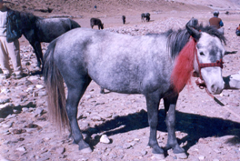
| Spiti ponies are high altitude small and stocky breed of equines which are found in cold deserts. These animals are the lifeline of the snow clad terrain. They are mainly used for transportation of goods. The Department of surgery and radiology has pioneered the need based anesthesia work in these animals as they are frequently presented for castration and other musculoskeletal disorders. |
| "Recommendations for Anesthesia in Spiti ponies |
Groups | Induction | Maintenance |
| DKX (Group I) (n=14) | Xylazine @ 1.1 mg/kg BW, IV & Ketamine @ 2.2 mg/kg BW, IV. | Diazepam 25mg, Xylazine 250mg & Ketamine 500mg in 500 ml normal saline as continuous intravenous drip infusion @ 2.2 ml /kg/ hr. |
| GKX (Group II) (n=14) | Xylazine @ 1.1 mg/kg BW, IV & Ketamine @ 2.2 mg/kg BW, IV. | Guaifenesin 25g, Xylazine 250mg & Ketamine 500 mg in 500 ml normal saline as continuous intravenous drip infusion @ 2.2 ml /kg/ hr. |
Anesthesia in Canine
Anesthesia in Canine |

Arthritis in yak
Arthritis in yak |
The fresh autogenous synovial fluid transfusion into arthritic animals has a positive therapeutic effect in decreasing lameness, increasing the joint flexion, relative viscosity and mucin precipitate quality and helps in early recovery from traumatic aseptic arthritis.